
Neurological Research: Fetal Tissue Brain Organoids Redefining Drug Research
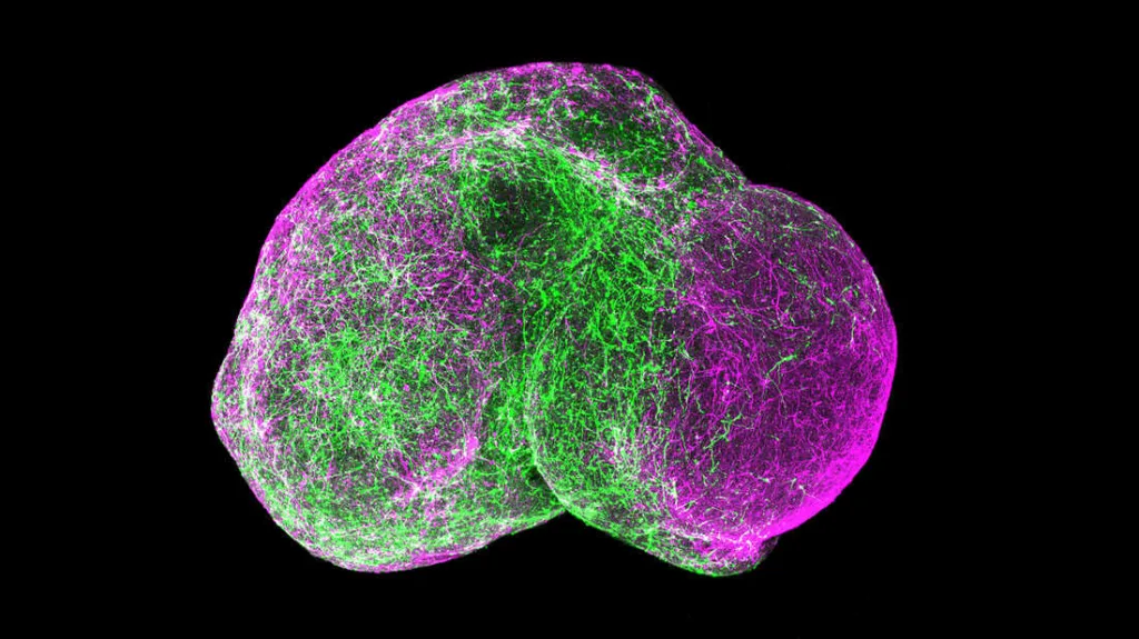
Neurological Research, The successful creation of three-dimensional mini-brains from human embryonic brain tissue is a significant development in neurological research. These microscopic organs, which are about the size of a rice grain, have self-organizing properties, opening up a previously unheard-of window into the complexities of brain development. Led by Dutch researchers, this innovation holds promising prospects for understanding and treating diseases linked to brain development, notably deadly brain tumors.


Revolutionizing Research Methods
The conventional approaches to modeling healthy tissue and diseases in labs involve cell lines, laboratory animals, and 3D mini-organs. These organoids, developed in 2011, replicate the functions of organs with remarkable accuracy. Unlike their predecessors, the latest brain organoids were created directly from human fetal brain tissue, a significant departure from the previous method of using individual cells.
Intricate Brain Organoid Development
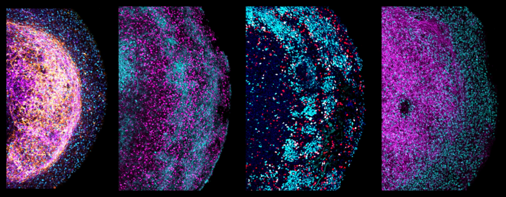
Published in the renowned journal Cell, the study from the Princess Máxima Center for Pediatric Oncology and the Hubrecht Institute reveals that brain organoids from fetal tissue present a leap forward. The team, led by Dr. Delilah Hendriks, Prof. Dr. Hans Clevers, and Dr. Benedetta Artegiani, observed that utilizing small pieces of fetal brain tissue allowed the organoids to self-organize. These miniature brains maintained characteristics specific to the region of the brain from which the tissue originated.


Unlocking the Potential of Brain Organoids
The significance of the extracellular matrix, the ‘scaffolding’ around cells, was highlighted in the study. Proteins from the whole pieces of brain tissue produced this matrix, contributing to the self-organization of the organoids. These brain organoids, resembling a complex 3D structure, contained various types of brain cells, including outer radial glia—a key cell type in human brain development.
Applications in Cancer Research
Beyond unraveling the mysteries of brain development, the researchers explored the potential of these organoids in modeling brain cancer. Using CRISPR-Cas9, they introduced faults in the TP53 gene, a known cancer gene, leading to the observation that the mutant cells acquired a growth advantage, a characteristic of cancer cells. This breakthrough allows for the study of specific gene mutations in relation to existing cancer drugs.
Future Prospects and Ethical Considerations
The researchers emphasize the potential of these brain organoids derived from fetal tissue in advancing our understanding of human brain development.
The ability to grow and utilize these organoids from precious fetal tissue offers a unique opportunity for groundbreaking discoveries. Moving forward, the scientists plan to further explore the capabilities of these organoids and collaborate with bioethicists to guide their future development and applications.
Insights from Lead Researchers:
Dr. Benedetta Artegiani: “Our new, tissue-derived brain model allows us to gain a better understanding of how the developing brain regulates the identity of cells.”
Dr. Delilah Hendriks: “Being able to keep growing and using the brain organoids from fetal tissue also means that we can learn as much as possible from such precious material.”
Prof. Dr. Hans Clevers: “It’s really exciting that we’ve now been able to jump that hurdle as well.”


